Examples of use

Scenario 1
I have the PDB coordinate file of a VH-VL antibody and I want to generate CRD sequence variants fitting the original backbones.

N. B: This coordinate file can be downloaded directly from the PDB or generated using a prediction tool (e.g., ABodyBuilder2) from a VH-VL sequence.

Follow these steps:

  • Click on the 'Choose file' button. This will open a window prompting you to browse your machine to select a PDB format file.
  • Once a file has been selected, select the CDR regions for sequence sampling. By default, all CDR regions for both heavy and light chain regions are selected. You can deselect by unticking the checkboxes as necessary.
  • Next, enter the chain identifiers of two paired antibody heavy and light chains in your coordinate file. If entered wrong, this will default into an error. If in doubt, check your PDB file for the right identifiers.
  • Finally, you can set how many sequences you want AntiFold to sample for you and their diversity via the 'Temperature' parameter.
  • After hitting the 'Submit' button, your job will be placed in a queue until finished.
Scenario 2
I have the PDB identifier of a VH-VL antibody structure available on SAbDab and I want to generate CRD sequence variants fitting the original backbones.

Follow these steps:

  • In the text box, enter a valid PDB ID (e.g., 8gho) of a structure available in SAbDab. If in doubt that it is a valid PDB, go to SAbDab and you can search for it.
  • Once entered, select the CDR regions for sequence sampling. By default, all CDR regions for both heavy and light chain regions are selected. You can deselect by unticking the checkboxes as necessary.
  • Next, enter the corresponding heavy and light chain identifiers of paired antibody heavy and light chains in your coordinate file. If entered wrong, this will default into an error. If in doubt, check the SAbDab page for your entry.
  • Finally, you can set how many sequences you want AntiFold to sample for you and their diversity via the 'Temperature' parameter.
  • After hitting the 'Submit' button, your job will be placed in a queue until finished.
Scenario 3
I don't have a PDB coordinate file or the SAbDAb PDB identifier of a VH-VL antibody structure. I just want to test the web app.

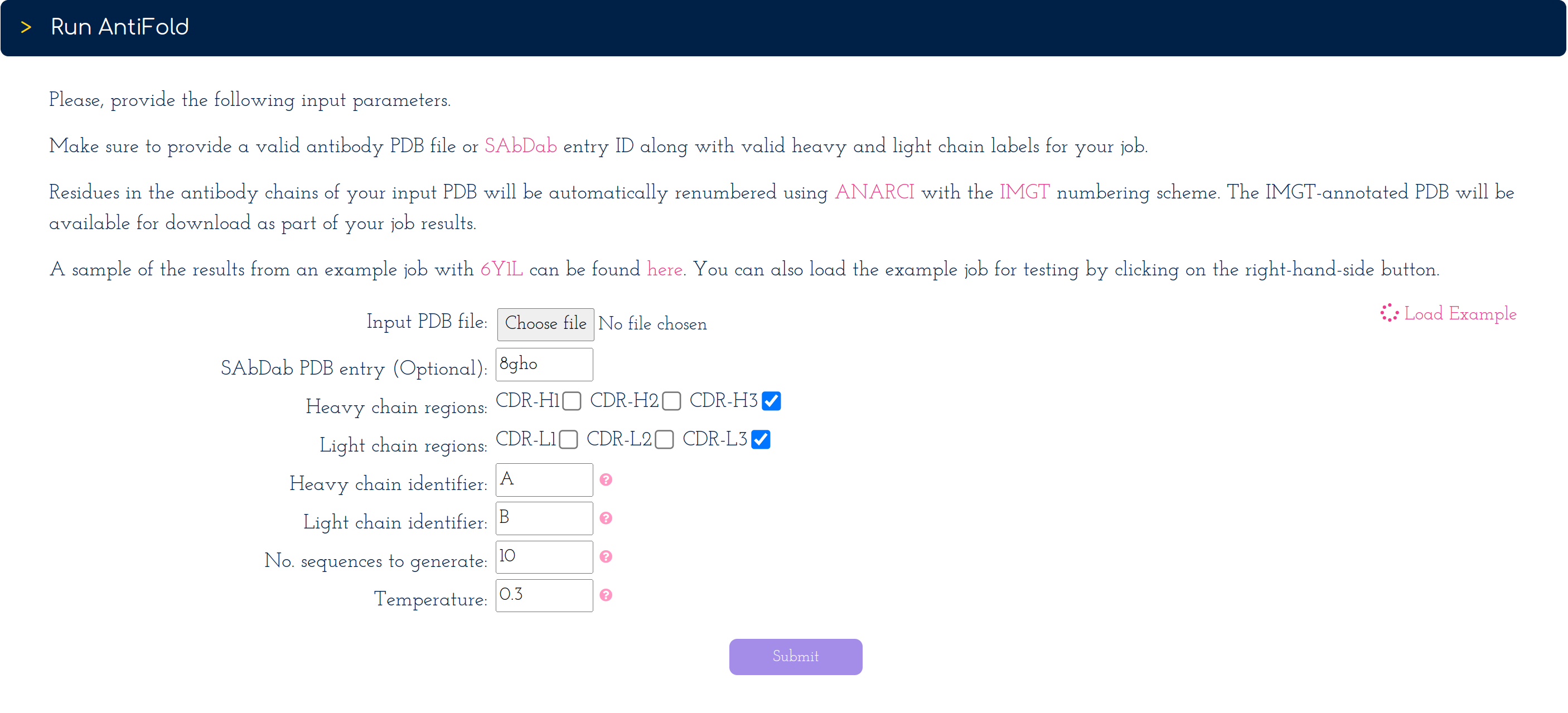
On the right-hand side of the submission form. You can click on the 'Load Example' button [See screenshot] to automatically load a PDB ID along with valid heavy and light chain identifiers.

As in scenarios 1 and 2, you can change all remaining parameters, if you wish.

After hitting the 'Submit' button, your job will be placed in a queue until finished.


Understanding the AntiFold web app results

Visualising the AntiFold perplexity per CDR residue

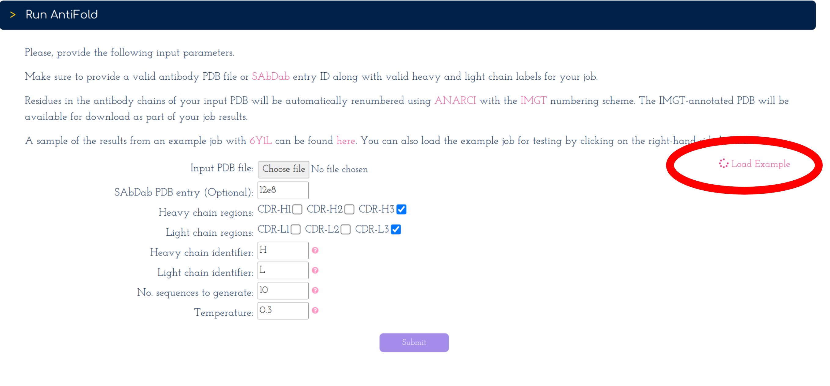
Perplexity at the residue level in the context of inverse folding is interpreted as the overall tolerance to mutations without altering the backbone structure.

The closer to perplexity equal one (1) a certain residue is, the less tolerant that residue position is to amino-acid variation. The opposite applies to high-perplexity positions.

The 'Structure Viewer' allows visualising AntiFold perplexity values of the CDR loops by clicking on the 'Perplexity' button under 'Color Options' [Right-hand-side screenshot].

To explore the perplexity CDR values for your input structure sequence the slider bar at the bottom of the viewer allows manipulation of the upper limit of the perplexity-encoded colour scale.

For instance, you can reveal which residues in the CDRs have the highest tolerance to mutation if they remain high in perplexity as you move the slider up.


Comparing AntiFold output CDR sequence variants against the original

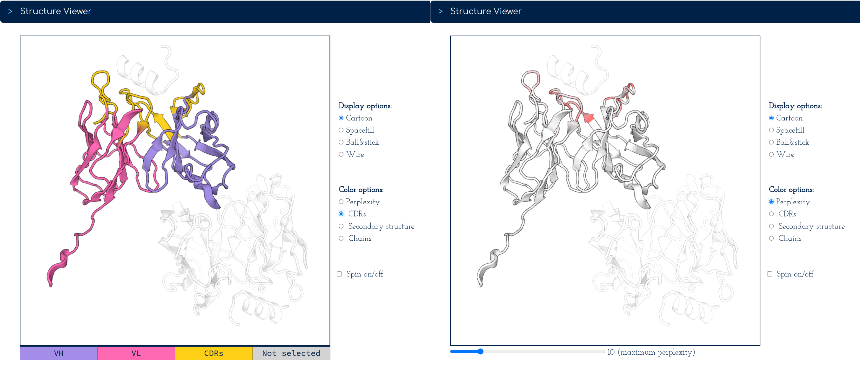
The original and variant sequences, labelled Seq N, are tabulated to highlight (in red) at which CDR loop positions point mutations fitting the original backbone are present.

CDR 1 to 3 loops are colour-coded and their IMGT numberings (obtained using ANARCI) are found in the top row of the table.

In the logo plots underneath the table, the inverse folding probabilities for all 20 essential amino acids are mapped to the height of their amino acid letter and its opacity, both ranging from 0 to 100%.

For example, for the heavy chain in the screenshot, at residue 105 in the CDR-3, Alanine (A) has a higher probability (~ 90%), in contrast to the original residue, Therionine (T).

Note that since we didn't select the CDR-1 and CDR-2 regions, all these residues are identical for the sequence variants Seq 1 to Seq 10 - as expected.

Downloads

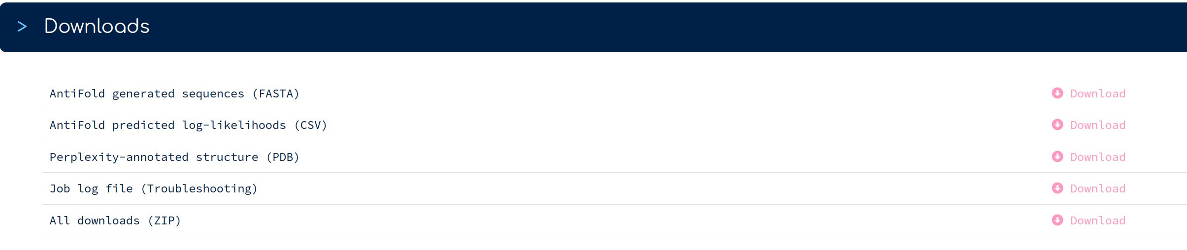
Here's a brief description of all file downloads, as listed:

  • Full-length Fv VH-VL sequences including the AntiFold CDR sequence variants tabulated in the 'Sequence Comparison' section can be downloaded in a FASTA file.
  • Amino-acid inverse folding probabilities used in logo plots are available in a CSV file.
  • In the PDB file download, beta-factor values are replaced with AntiFold perplexity values. Perplexities for the full Fv sequence can be found in the CSV file.
  • The log file download can be helpful in troubleshooting if your job fails.
  • The ZIP archive does feature all the above files, except the logo plots and other complementary files the web app uses to execute your job.

Contact

Feel free to contact us at opig <~at~> stats.ox.ac.uk for any issues, or general enquiries about the AntiFold web app.

For specific issues about the AnitFold Python code, you can open an issue directly on the AntiFold GitHub repo.

Magnus Haraldson Høie & Alissa Hummer et al. “AntiFold:
Improved antibody structure-based design using inverse folding” 2024. [ link]

Download BibTex Reference

Header image credit: David Goodsell